
Serving our New Jersey Community


How X-Rays Work
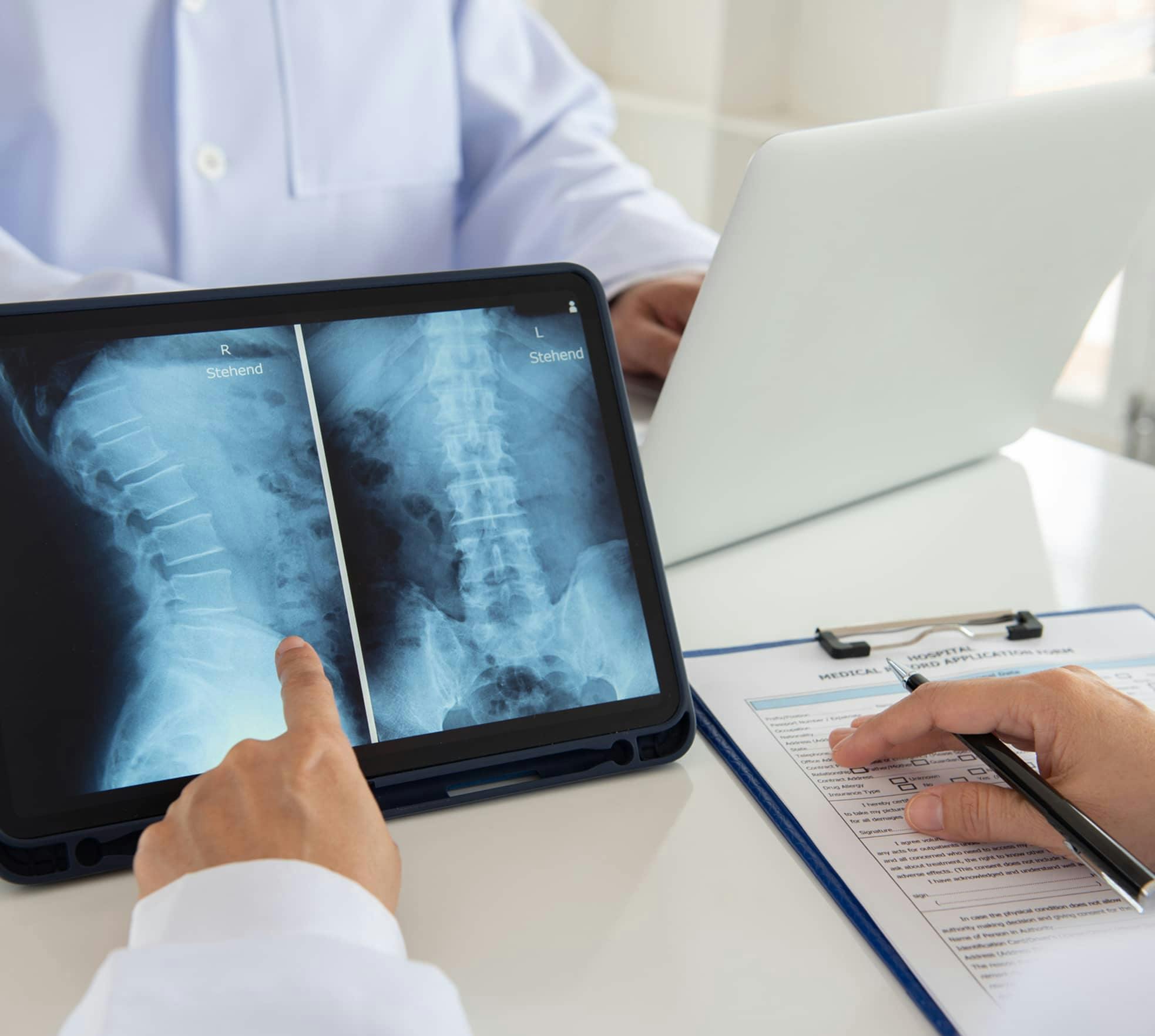
During an X-ray, low doses of invisible electromagnetic energy with short wavelengths pass through your body, creating a shadowy image on a film or screen. Bones, being dense, absorb X-rays effectively and appear as white on the film, while tissues display as darker areas.
The X-Ray Procedure
During an X-ray examination, you'll lie on a table with an adjustable X-ray machine positioned above you. The procedure is painless, and you won't feel anything as the X-rays pass through your body. To ensure clarity, it's essential to remain still while the image is captured. In cases of scoliosis and kyphosis, X-rays are often taken with the patient standing.
Safety and Radiation
X-ray exposure time is extremely brief, and modern equipment ensures minimal radiation doses. However, X-rays are typically avoided during pregnancy, and a lead shield should be used to protect you from X-ray beams.