
The Center for the Functional Restoration of the Spine (CFRS), with locations in Shrewsbury, Toms River, and Edison, New Jersey, specializes in accurately diagnosing and effectively treating painful back conditions.
Orthopaedic spine specialists, Steve Paragioudakis, MD, and Marc Menkowitz, MD, lead our team of dedicated professionals whose goal is to provide you the most effective care possible in a comfortable and welcoming environment that’s always patient-focused.
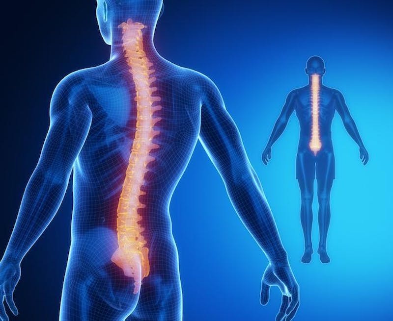
Here, we give you a closer look at the diagnostic studies your CFRS specialist may use to uncover the cause of your back pain.
Diagnosing your back pain
Based on a careful evaluation that includes a physical exam and thorough discussion of your symptoms, we may recommend one or more of the following diagnostic tests to accurately identify the source of your pain.
Traditional X-rays
Familiar to most people, plain X-rays offer a quick diagnostic view of spinal issues such as:
- Degenerative disc disease
- Scoliosis
- Kyphosis
- Stenosis (narrowing of the spinal canal)
Because traditional X-rays only highlight bones, they are not helpful in diagnosing soft tissue injuries, such as muscle strains or ligament sprains.
CT scan
A computerized tomography scan (CT scan) combines X-rays with computer technology to offer a detailed three-dimensional view of your spinal structures. Sometimes combined with other studies such as a myelogram or discogram for the most accurate results, a CT scan may last from 15 minutes to an hour.
CT scans are painless and less noisy than an MRI. You won’t, however, be able to eat solid food for three hours before the study.
Magnetic resonance imaging (MRI)
The MRI provides detailed images of bones as well as soft tissue structures. It does take longer than a CT scan, up to 90 minutes, and is generally more expensive, so it’s not typically the first diagnostic choice.
An MRI may be necessary, however, to provide an accurate view of a vertebral infection, spinal tumor, or other condition. The study is painless but somewhat noisy. Individuals with pacemakers, aneurysm clips, or other non-removable metallic devices cannot have an MRI.
Bone scan
A bone scan is an imaging study we use to evaluate spinal tumors, infection, or fractures more thoroughly than is possible with a traditional X-ray.
During the study, we inject a radioactive chemical into your bloodstream that adheres to damaged portions of your spine. These regions show up as darkened areas on the image provided by a gamma camera.
Radiation levels in the chemical used during a bone scan are lower than those found in traditional X-rays. You may feel mild discomfort briefly during the injection, but the study is otherwise painless.
Discogram
A discogram includes traditional X-rays but is often combined with a CT scan for better accuracy. During the study, we inject a special dye into one or more spinal discs to help locate those responsible for your discomfort. A herniated or ruptured disc leaks dye into the surrounding tissue, which is visible on X-ray.
Use of IV sedation during a discogram means you’ll need a ride home after the procedure and should rest overnight.
Myelogram
During a myelogram, we inject dye into your spinal column to reveal the flow of fluid through your spinal column. X-rays may be combined with a CT scan to present the most accurate view. Decreased or irregular flow of spinal fluid may indicate nerve pressure related to a herniated disc or bone spur. Rarely, it may also signal a spinal tumor.
For an accurate diagnosis and treatment you can rely on to relieve your back pain, schedule an evaluation at CFRS today. Call our office or request an appointment online.